December 29, 2024 by 0 Comments

What is Angioplasty?

Angioplasty is a medical procedure used to open narrowed or blocked blood vessels, particularly in the heart. This method is primarily employed to treat cardiovascular diseases, such as coronary artery disease (CAD).

  1. History

Angioplasty was first introduced in 1977 by Dr. André Gruntzig, a cardiologist. Since then, this procedure has been widely used in the treatment of vascular problems, with significant advancements in technology and techniques.

  1. Procedure of Angioplasty

Angioplasty is typically performed as follows:

  • Preparation: The patient is usually given local anesthesia and may also receive sedation.

  • Accessing the Vessels: A catheter (a thin, flexible tube) is inserted through a blood vessel in the arm or leg and guided towards the coronary arteries.

  • Imaging: Using X-rays and a contrast agent, the doctor can visualize the condition of the blood vessels.

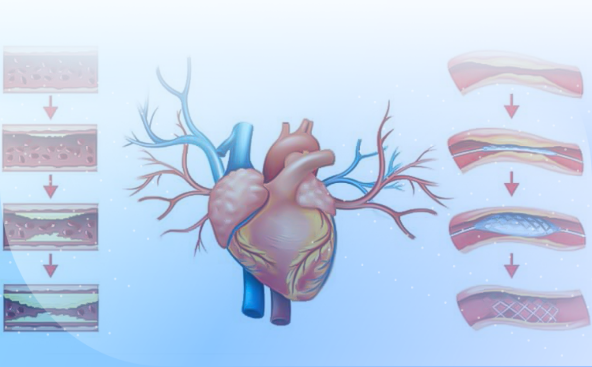
  • Opening the Vessels: A small balloon at the end of the catheter reaches the site of narrowing or blockage and inflates to open the vessels. In many cases, a stent (a mesh tube) is also placed at the site of narrowing to prevent re-narrowing.

  1. Advantages and Disadvantages

Advantages:

  • Improved blood flow to the heart
  • Reduced symptoms of heart disease, such as chest pain
  • Enhanced quality of life

Disadvantages:

  • Side effects such as bleeding, infection, or allergic reactions to the contrast agent
  • Possibility of re-narrowing of the blood vessels
  1. Post-Angioplasty Care

Patients typically require special care after undergoing angioplasty. This includes taking blood-thinning medications, making lifestyle changes, and having regular follow-ups with their doctor.

  1. Conclusion

Angioplasty is an effective procedure for treating vascular issues that can help improve the quality of life for patients. However, like any medical procedure, it requires careful evaluation and consultation with a specialist physician.

December 29, 2024 by 0 Comments

Hypertension Treatment: A Comprehensive Guide

Hypertension (high blood pressure) is a common health issue that can lead to serious complications such as heart disease, stroke, and kidney disease. Treatment for hypertension involves lifestyle changes and, if necessary, medication. In this article, we will explore various methods for treating hypertension.

  1. Diagnosis of High Blood Pressure

Blood pressure is divided into two numbers: systolic blood pressure (the upper number) and diastolic blood pressure (the lower number). Normal blood pressure is typically less than 120/80 mmHg. High blood pressure is classified into two categories:

  • Stage 1: Systolic blood pressure between 130 and 139 or diastolic blood pressure between 80 and 89.
  • Stage 2: Systolic blood pressure of 140 or higher or diastolic blood pressure of 90 or higher.
  1. Lifestyle Changes

Lifestyle changes can significantly impact blood pressure control. These changes include:

  • Healthy Diet:

    • Reducing sodium intake to less than 2,300 mg per day.
    • Increasing the consumption of fruits, vegetables, whole grains, and lean proteins.
    • Following the DASH diet (Dietary Approaches to Stop Hypertension), which emphasizes the intake of nutrients like potassium, calcium, and magnesium.
  • Physical Activity:

    • Engaging in at least 150 minutes of moderate exercise per week, such as walking, cycling, or swimming.
  • Weight Management:

    • Losing weight in individuals who are overweight can help reduce blood pressure.
  • Stress Management:

    • Utilizing stress-reduction techniques such as meditation, yoga, or breathing exercises.
  • Quitting Smoking and Reducing Alcohol Consumption:

    • Smoking and alcohol consumption can increase blood pressure. Quitting these habits can improve heart health.
  1. Medications for Hypertension

If lifestyle changes are insufficient, a doctor may prescribe medications. Various types of medications are available for treating hypertension:

  • Diuretics: These medications help reduce blood volume.
  • Beta-blockers: These medications lower heart rate and reduce blood pressure.
  • ACE Inhibitors: These medications help dilate blood vessels and lower blood pressure.
  • Calcium Channel Blockers: These medications help reduce vascular contraction and lower blood pressure.
  • Angiotensin II Receptor Blockers: These medications also assist in dilating blood vessels.
  1. Blood Pressure Monitoring

Regular monitoring of blood pressure is essential to ensure effective control. Patients can measure their blood pressure at home using digital blood pressure monitors and share the results with their doctor.

  1. Conclusion

Managing high blood pressure requires a comprehensive approach that includes lifestyle changes and, if necessary, medication. By following appropriate strategies and consulting with a physician, blood pressure can be reduced to a healthy level, preventing serious complications.

December 29, 2024 by 0 Comments

ECG Interpretation: A Comprehensive Guide

Electrocardiography (ECG) is a key tool in the diagnosis and assessment of heart diseases. This test records the electrical activity of the heart, providing important information about heart rhythm, size and position of the heart, and the presence of any functional disorders. In this article, we will explore how to interpret an ECG and key points to consider.

  1. Structure of the ECG

The ECG is recorded as a curve on paper or a display and includes several waves and intervals:

  • P Wave: Represents atrial contraction (sinoatrial).
  • QRS Complex: Represents ventricular contraction.
  • T Wave: Represents the return of the ventricles to a resting state.
  • PR Interval: The time between the start of atrial contraction and the start of ventricular contraction.
  • QT Interval: The time of ventricular contraction and the return to a resting state.
  1. Steps in ECG Interpretation

Interpreting an ECG involves several key steps:

a) Assessing Heart Rhythm

  • Regular or Irregular: Determine whether the heart rate is regular or irregular. A normal heart rhythm typically ranges from 60 to 100 beats per minute.
  • Source of Rhythm: Identify whether the rhythm originates from the sinoatrial (SA) node. The presence of a P wave before each QRS indicates a sinus rhythm.

b) Measuring Heart Rate

  • Heart Rate: Calculate the number of beats in one minute. To calculate, count the number of QRS complexes in a 10-second period and multiply by 6.

c) Analyzing Waves

  • P Wave: Check if a P wave exists before each QRS and whether its shape is normal.
  • QRS Complex: Measure the duration of the QRS (it should be less than 0.12 seconds) and assess its shape.
  • T Wave: Examine the shape and height of the T wave. Changes in the T wave may indicate issues such as cardiac ischemia.

d) Measuring Intervals

  • PR Interval: Should be between 0.12 and 0.20 seconds. An increase in this interval may indicate AV block.
  • QT Interval: Should be less than 0.44 seconds. Prolongation of this interval can increase the risk of arrhythmias.
  1. Identifying Disorders

ECG interpretation can help identify various disorders:

  • Coronary Artery Disease: May manifest with changes in the T wave or ST segment.
  • Congenital Heart Diseases: Can be identified by changes in the shape and size of waves and intervals.
  • Arrhythmias: Such as atrial fibrillation or ventricular tachycardias, which are characterized by changes in heart rhythm.
  • Electrolyte Disorders: Changes in waves and intervals may indicate electrolyte imbalances such as hypokalemia or hyperkalemia.
  1. Conclusion

ECG interpretation is a key skill in medicine that requires training and practice. By mastering the steps of interpretation and recognizing various disorders, physicians can aid in the accurate diagnosis and effective treatment of heart diseases.

November 9, 2024 by 0 Comments

Echocardiography: A Comprehensive Guide

chocardiography (Ultrasound Echocardiography) is a non-invasive imaging technique used to assess the structure and function of the heart. This method uses sound waves to create images of the heart and blood vessels, providing important information about the heart’s condition. In this article, we will explore the types of echocardiography, how it is performed, its applications, and the benefits of this method.

  1. Types of Echocardiography

Echocardiography is divided into several types:

  • Transthoracic Echocardiography (TTE): This is the most common type of echocardiography, performed by placing a probe on the chest. This method is suitable for a general assessment of the heart and blood vessels.

  • Transesophageal Echocardiography (TEE): In this method, the probe is inserted into the esophagus to obtain more detailed and clearer images of the heart and blood vessels. This method is typically used when TTE does not provide sufficient results.

  • Stress Echocardiography: This type of echocardiography helps assess heart function under stress conditions (usually through exercise or medication). This method can help identify coronary artery diseases.

  • Three-Dimensional Echocardiography: This type of echocardiography creates three-dimensional images of the heart, helping physicians better visualize cardiac structures.

  1. How Echocardiography is Performed

Echocardiography is usually performed as follows:

  • Preparation: The patient typically lies down. Anesthesia is not required, and the patient can remain awake during the procedure.

  • Probe Placement: The physician applies a special gel to the skin of the patient’s chest and places the probe on it. This gel aids in the transmission of sound waves.

  • Imaging: The probe sends sound waves to the heart and receives the echoes of these waves. These echoes are converted into images of the heart.

  • Image Analysis: The physician analyzes the obtained images and examines information related to the size, shape, and function of the heart.

  1. Applications of Echocardiography

Echocardiography is used for the diagnosis and assessment of various heart diseases, including:

  • Valvular Diseases: Evaluating the function of heart valves and identifying valve insufficiency or stenosis.

  • Cardiac Muscle Diseases: Such as cardiomyopathy and heart failure.

  • Coronary Artery Diseases: Identifying problems caused by arterial blockages.

  • Congenital Heart Disorders: Assessing the structure of the heart in newborns and children.

  • Preoperative Care: Evaluating the heart’s condition before cardiac surgeries.

  1. Benefits of Echocardiography
  • Non-invasive: Echocardiography is a non-invasive method that does not require surgery or entry into the body.

  • No Side Effects: This method has no specific side effects due to the use of sound waves and the absence of X-rays.

  • High Accuracy: Echocardiography can provide accurate information about the structure and function of the heart.

  • Real-time Capability: This method allows for the observation of heart movements in real time.

  1. Conclusion

Echocardiography is a key tool in the diagnosis and assessment of heart diseases, providing valuable information about the function and structure of the heart. Given its non-invasive nature and high accuracy, this method is recognized as one of the primary techniques in cardiovascular medicine.